Latvijas kvalifikāciju datubāze
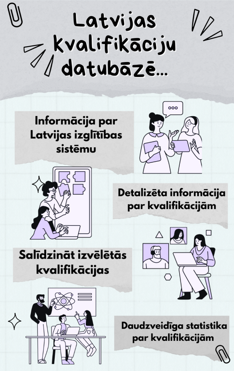
Latvijas kvalifikāciju datubāze (LKD) ir Latvijā unikāla datubāze, kurā ir apkopotas Latvijas izglītības sistēmā iegūstamās kvalifikācijas. LKD kvalifikācija ir zināšanas, prasmes un kompetences, ko apliecina izglītības dokuments.
Latvijas kvalifikāciju datubāzē kvalifikācijas ir strukturētas atbilstoši Latvijas kvalifikāciju ietvarstruktūras (LKI) un Eiropas kvalifikācijas ietvarstruktūras (EKI) astoņiem līmeņiem:
Made with Visme Presentation Maker
Video: Kā un kādēļ izmantot Latvijas kvalifikāciju datubāzi?